for Health Care Providers
HIV-Related Cancers
Back to: Image Library | HIV-Related Cancers Images
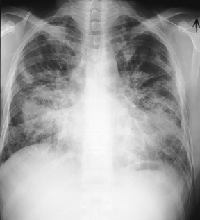
Kaposi sarcoma, pulmonary: chest X ray
Description
Chest radiograph of an HIV-infected man, CD4 count of < 100 cells/ µ L, demonstrating bilateral, predominantly middle and lower lung zone abnormalities in a central distribution. The abnormalities include a central coalescent pattern with smaller, nodular opacities best seen in the periphery of the upper lung zones. Kerley B lines can be appreciated at the bases. This constellation of findings in an HIV-infected man is suggestive of pulmonary Kaposi sarcoma (KS). The diagnosis was confirmed by bronchoscopic visualization of the characteristic KS lesions seen throughout the tracheobronchial tree.
Credits
Laurence Huang, MD, University of California San Francisco