


for Health Care Providers
Radiographic Studies
Back to: Image Library | Radiographic
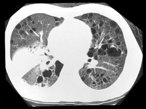
Pneumocystis jiroveci (formerly carinii ) pneumonia
Description
Computed tomography scan of the chest of an HIV-infected patient, CD4 count of < 200 cells/ µ L, with multiple, bilateral cysts secondary to Pneumocystis jiroveci as well as a focal alveolar consolidation secondary to Streptococcus pneumoniae .
Credits
Laurence Huang, MD, University of California San Francisco


