


for Health Care Providers
Radiographic Studies
Back to: Image Library | Radiographic
Pneumococcal pneumonia: bacteremic, with Pneumocystis jiroveci (formerly carinii )
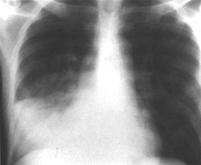
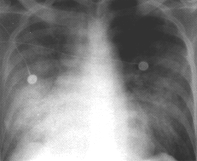
Description
Figure 1: Chest radiograph of an HIV-infected patient with bacteremic pneumococcal pneumonia showing right middle lobe consolidation.
Figure 2: Repeat chest radiograph 8 days later when the patient developed worsening shortness of breath and hypoxemia. The film shows worsening bilateral heterogenous infiltrates due to
Pneumocystis jiroveci
pneumonia.
Credits
Charles L. Daley, MD, University of California San Francisco


