


for Health Care Providers
Pneumocystis jiroveci (formerly carinii)
Back to: Image Library | Pneumocystis jirovecii
Pneumocystis jiroveci : HRCT scan in a patient with a normal chest X ray
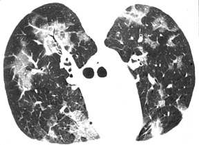
Description
High-resolution computed tomograph (HRCT) scan of the chest of an HIV-infected patient, CD4 count of < 200 cells/ µ L, who had a normal chest radiograph. Bilateral patchy areas of ground glass opacity are suggestive of Pneumocystis jiroveci pneumonia, which was microscopically confirmed by examination of induced sputum.
Credits
Laurence Huang, MD, University of California San Francisco


